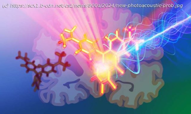
To understand the brain better, we need new methods to observe its activity. That is at the heart of a molecular engineering project, spearheaded by two research groups at the European Molecular Biology Laboratory (EMBL), that has resulted in a novel approach to create photoacoustic probes for neuroscience applications. The findings were published in the Journal of the American Chemical Society.
To understand the brain better, we need new methods to observe its activity. That is at the heart of a molecular engineering project, spearheaded by two research groups at the European Molecular Biology Laboratory (EMBL), that has resulted in a novel approach to create photoacoustic probes for neuroscience applications. The findings were published in the Journal of the American Chemical Society.
« Photoacoustics offer a way to capture imagery of an entire mouse brain, but we just lacked the right probes to visualize a neuron’s activity », said Robert Prevedel, an EMBL group leader and a senior author on this paper. To overcome this technological challenge, he worked with Claire Deo, another EMBL group leader and also a senior author on the paper. She and her team specialize in chemical engineering.
« We have been able to show that we can actually label neurons in specific brain areas with probes bright enough to be detected by our customized photoacoustic microscope », Prevedel said.
Scientists can learn more about biological processes by tracking certain chemicals, such as ions or biomolecules. Photoacoustic probes can act as « reporters » for hard-to-detect chemicals by binding to them specifically. The probes can then absorb light when excited by lasers and emit sound waves that can be detected by specialized imaging equipment.
Home
United States
USA — IT New photoacoustic probes enable deep brain tissue imaging, with potential to report...


